Gentle Imaging of Lipid Bilayers with Encased Cantilevers

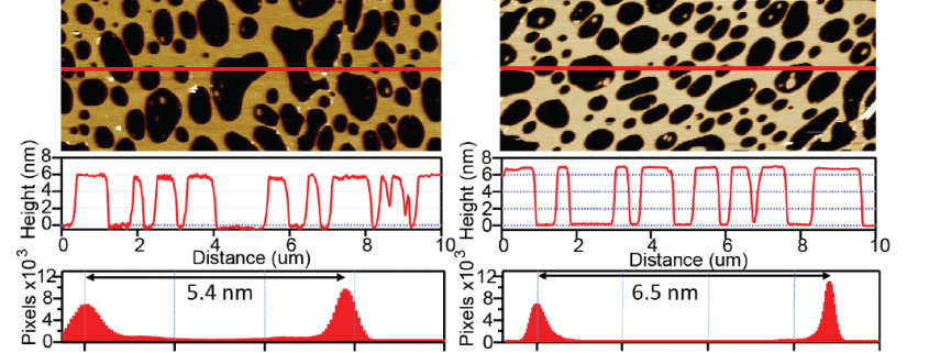
High viscous damping has limited the AFM’s utility for high resolution imaging of soft materials in solution resulting in sample deformation or damage. Lipid bilayers readily deform under the force applied by the AFM tip. Scuba Probe’s encased cantilevers offer gentler imaging conditions as shown here imaging supported DPPC lipid bilayers (L-αdipalmitoyl-phosphatidylcholine) that were prepared using a Langmuir-Blodgett trough, and transferred onto mica in an aqueous buffer. The softest imaging conditions achieved with (a) conventional silicon nitride cantilevers (Hydra Cantilevers, Applied Nanosciences Inc.) results in a bilayer height of 5.4 nm. In contrast, (b) encased cantilevers reveal a bilayer thickness of 6.5 nm. This bilayer thickness exceeds the highest previously reported thickness using AFM, clearly demonstrating the gentle imaging enabled by the reduced viscous damping.