High Resolution Imaging of DNA Origami with Encased Cantilevers
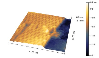
 Rationally designed nanoscale materials hold great promise for a wide variety of applications including sustainable energy, photonics, biophysics, and medicine. In particular, DNA origami offers precise organization of nucleic acids that enables the construction of both 2D and 3D structures. Specific functionalities can be directly designed into the structure or they can be used directly or used as a scaffold to template new materials. To visualize the detailed structure of DNA origami assemblies, gentle imaging conditions are required. Here we show a high resolution tapping mode AFM image of a 2D DNA origami rectangle in a TAE buffer obtained using an encased cantilever. Individual staples and crossover points in the structure can be easily identified while imaging in liquid.
A special thank you to Nicholas Stephanopoulos and his group from Arizona State University for providing samples.
Ultra-Low Force Noise and Stability with Electrostatic Actuation

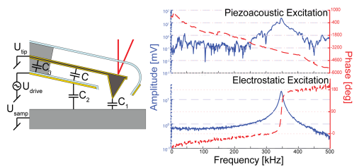
Encased cantilevers are novel force sensors that overcome major limitations of liquid scanning probe microscopy. By trapping air inside an encasement around the cantilever, they provide low damping and maintain high resonance frequencies for exquisitely low tip–sample interaction forces even when immersed in a viscous fluid. Quantitative measurements of stiffness, energy dissipation and tip–sample interactions using dynamic force sensors remain challenging due to spurious resonances of the system. We demonstrate for the first time electrostatic actuation with a built-in electrode. Solely actuating the cantilever results in a frequency response free of spurious peaks. We analyze static, harmonic, and sub-harmonic actuation modes. Sub-harmonic mode results in stable amplitudes unaffected by potential offsets or fluctuations of the electrical surface potential. We present a simple plate capacitor model to describe the electrostatic actuation. The predicted deflection and amplitudes match experimental results within a few percent. Consequently, target amplitudes can be set by the drive voltage without requiring calibration of optical lever sensitivity. Furthermore, the excitation bandwidth outperforms most other excitation methods. Compatible with any instrument using optical beam deflection detection electrostatic actuation in encased cantilevers combines ultra-low force noise with clean and stable excitation well-suited for quantitative measurements in liquid, compatible with air, or vacuum environments.
DOI: 10.3762/bjnano.9.130
Elucidating Water Structure with Ultrasensitive Cantilevers
 The local hydration structure at the liquid/solid interface influences the function of adsorbed molecules that can range in complexity from a single ion to complex biomolecules. The low damping, high Q, and low force noise that are characteristic of encased cantilevers enable high resolution imaging combined with ultrasensitive force measurement as demonstrated in the high-resolution image shown in (a) that resolves the muscovite mica atomic lattice. Force measurements on the same surface are shown in (b) and schematically in (c) further illustrate the sensitivity of the encased levers. Three distinct oscillations are observed at the mica surface, representing solvent-molecule layers. To date, three structured water layers have been observed in AFM over assembled biomolecules, but only two water layers have been observed on mica. Moreover, the single force curve measured in (b) was taken in only one second and was not averaged, demonstrating one of the most sensitive AFM measurements of water structure to date. Overall, the increased sensitivity offered by encased cantilevers could push AFM to resolve 2D and 3D hydration layers on surfaces in liquid.
Gentle Imaging of Lipid Bilayers with Encased Cantilevers
 High viscous damping has limited the AFM’s utility for high resolution imaging of soft materials in solution resulting in sample deformation or damage. Lipid bilayers readily deform under the force applied by the AFM tip. Scuba Probe’s encased cantilevers offer gentler imaging conditions as shown here imaging supported DPPC lipid bilayers (L-αdipalmitoyl-phosphatidylcholine) that were prepared using a Langmuir-Blodgett trough, and transferred onto mica in an aqueous buffer. The softest imaging conditions achieved with (a) conventional silicon nitride cantilevers (Hydra Cantilevers, Applied Nanosciences Inc.) results in a bilayer height of 5.4 nm. In contrast, (b) encased cantilevers reveal a bilayer thickness of 6.5 nm. This bilayer thickness exceeds the highest previously reported thickness using AFM, clearly demonstrating the gentle imaging enabled by the reduced viscous damping.
Encased Cantilevers Make Quantitative Mass Sensing Simple
 Cantilever based mass sensors detect minute amounts of material and have the potential to significantly impact biological and chemical diagnostics. Added mass to the cantilever is directly measured through a resonance frequency shift, however the high damping-low Q-factor environment of liquids makes measurements of resonance frequency challenging. The low damping of encased cantilevers enables in-situ mass sensing in liquids that surpasses conventional cantilever based mass sensing, where the uncertainty of the location of the added mass, and the indirect measurement of stress induced bending require various assumptions or models to extract the exact value of the added mass. With encased cantilevers, (a) functionalization and binding of an analyte occur only at the tip and (b) a distinct shift in frequency is easily observed from two consecutive attachment events using 250 nm gold particles. The volume of the gold particles (c) shown in high-angle annular dark field electron microscopy was estimated by projected areas and assuming a spherical form factor (volume estimation in d, green histogram). True mass sensing using encased cantilevers is shown in d (red histogram), where an average mass of 168 fg/nanoparticle is measured. With the potential to reach even attogram sensitivities, encased cantilevers can be used as a sensitive mass sensor for a variety of applications in solution.
Scanning Electrochemical Microscopy using Insulated Cantilevers
 Electrochemical processes are central to corrosion, energy storage, catalysis, and biology. Beyond the topography that is measured in atomic force microscopy, scanning electrochemical microscopy (SECM) measures reaction rates and electrochemically generated species. Here, we demonstrate the capability of Scuba Probe Technologies insulated cantilevers to measure electroactive species being generated at the sample surface. By probing a micro electrode array in hexaammineruthenium (III) chloride operating in sample generation-tip collection mode, we demonstrate that electrical currents on the order of a few pA can be observed. Correlating current (top) with topography (bottom) allows for identification of both active and inactive electrodes.

